

This week, regular random post service has been suspended for something all-the-more exciting. On Sunday this week, I managed to get an MRI for my ankle through Advanced Imaging Westmead at Westmead Private Hospital. The service was rendered through referral, under Medicare coverage, so I didn’t have to pay, which is great.
My First MRI
Having never had an MRI before, it was rather fascinating to try and get a grasp of just how MRI works. Unfortunately, I still don’t have a clear idea, but somehow it involves using coiled superconducting electromagnets to generate fields which are pulsed in order to observe the radio frequency emissions from decaying hydrogen atoms (or something like that). Being an engineer, and an RF person, I had some appreciation of how “serious” such a device would be.
One obvious result was that you couldn’t have magnets or metal near the machine. Before entering the MRI suite, I had to remove my belt and empty my pockets. Because of the high currents and switching frequency being within audible frequencies, I was warned about very very loud noises described as “knocking”, or “beeping”. That’s expected from the minute movements of the coils under switched conditions (same reason why some power packs “hum”).
My ankle was strapped in, I was moved into the tube. The operator put earmuffs over my ears, and closed the door. She remotely commanded the imaging sequences from outside, and the machine’s LCD turned on and off between each batch. The machine didn’t so much knock or beep, but sounded like the over-the-horizon radar that we hear on shortwave quite often. Bzzt-bzzt-bzzt-bzzt-bzzt. Brrp-brrp-brrp-brrp-brrp-brrp-brrp-brrp. It was rhythmic, except for when we got to the end of a sequence and it would pause for a bit before commencing the next. I’d imagine being next to a radar machine might be similar. I was actually quite mentally comfortable, although wondering just how much energy was going into these coils as I watched the downlights flicker quite severely. My leg though, wasn’t so happy. It was cramping up. But I held still as I could and before long, it was over.
In short, nothing to be afraid of, and in fact, an engineering and medical marvel to think that there’s no known damage from this. No radiation involved (as no contrast was used). I might have experienced some Peripheral Nerve Stimulation (PNS) I believe, as at one imaging sequence in the scan, my toes uncontrollably tingled and moved slightly.
The Equipment
It was only after the scan that I took note of what equipment was used. It was a Siemens Magnetom Verio Tim+Dot MRI system. It’s quite an advanced machine, being a 3 Tesla (3T) model, where older units were 1.5T. Higher field strengths can mean quicker imaging, so that’s a lovely sign. The room it was housed in was a faraday cage to ensure radio frequency interference from outside is minimised (as I can imagine the resonance signal would be extremely weak). The windows were covered in dark film (likely conductive, like the heating film on modern car windscreens), and the door edges were covered in springy metal fingers to make a good contact seal around the door frame.
Outside the imaging suite, the command was all done on Fujitsu workstations. I’m not sure what they were running, but it was a dedicated full-screen GUI with two workstations. Output could (probably) be sent to DICOM files and burnt to DVD (there were piles of Verbatim DVD’s there), or sent to their Agfa DryStar 5500 film printer. They had boxes of Ricoh film there as well.
I would definitely say that I would have preferred a DICOM disc, but the service only provided 12 pieces of film and I didn’t bother asking. Maybe the referring doctor isn’t readily equipped to display and handle DICOM files on DVD, or maybe it’s an additional cost extra not covered by Medicare. Film has its benefits too with long term storage, but for my own tinkering, it’s a bit of a mess.
Working with Imagery
I don’t think I really have to say it, but I’m not qualified in any way to read or interpret any medical diagnostic imagery. I’m not trained in how the equipment works, what all the settings mean, etc. But I’m always interested in the data.
The first thing I had to do was to acquire the data from film. As I don’t have an “x-ray film” scanner, I had to do something different. I tried using a regular flat-bed, but the light doesn’t really have enough oomph to go through the film twice, and we get a lot of surface grain being picked up.
So I improvised a solution involving a Solar Panel, three flash heads (one on the body, two by remote optical trigger) and my DSLR. I used my newly acquired Micro-Nikkor AF-S 105mm f/2.8G IF-ED VR and my D3200 to do the shooting.

The white tedlar backing of the cheap solar panel provides a nice “diffusing” screen. The frame elevates the imagery off the tedlar by about 20mm, and cello tape is used to hold it in place. The flashes provide quick back-illumination making hand-held shooting viable.
Unfortunately, the lighting wasn’t 100% even, we did have a few bright spots and focus was impossible except for by manual focus. I still managed to eventually (after a lot of back and forth), get all 12 films photographed. Then I had to crop and deskew each one manually to get individual images, resize them and animate.
Of course, if I had the DICOM files, I wouldn’t have needed to do any of this. Photoshop can open DICOM files directly – so if your DICOM viewer on your disc doesn’t work, Photoshop definitely does!
I did think of using an LCD monitor as the backlight, but I decided against it due to early trials showing the pixels through the film (if it’s within the DOF of the lens settings), and it’s not really comfortably bright enough to be hand-held shot.
See my MRIs – Click for Animated!
In total, I had six imaging sequences performed. with 147 separate images printed to film. As I’m not suitably qualified to interpret the scans, I’ll try to note my observations (although they could be wrong). Image sequences prior to 3 were not printed, as they were likely used for probing and determining what area is of interest for imaging (kind of like the “pre-scan” feature of your scanner, to check the alignment and highlight the zone for further scanning).
Please note that the following files aren’t for diagnostic purposes – I have had to alter them slightly to redact some patient data, and compensate for “image acquisition limitations”. This involves clearing up shiny parts of the film, adjusting for uneven brightness (as best as I can) and filling in black for parts where the edges didn’t quite make it into the photo that I took.
As the animations range from 2-5Mb each, I’ve put some static images here. Click to see the animated version.

Image Sequence No. 3 – Scan focusing on bone, slices from the left side of the foot, through the ankle to the right side of the foot.
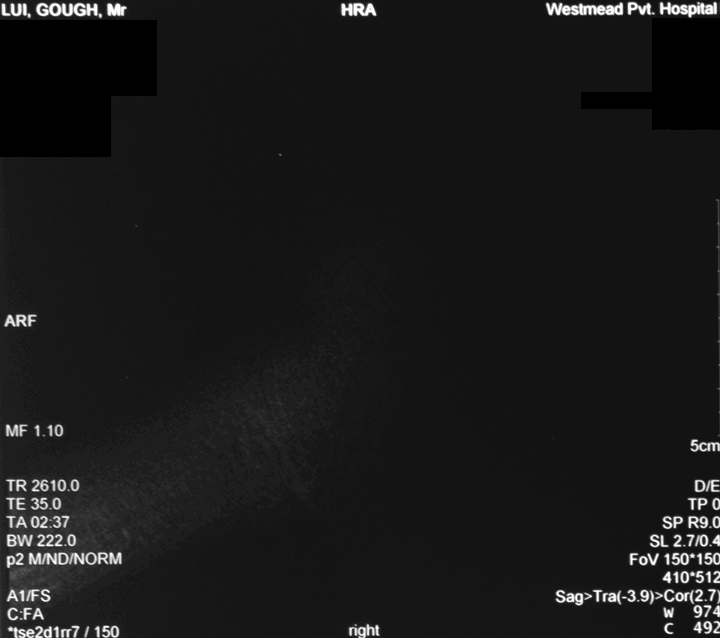
Image Sequence No. 4 – Scan focusing on soft tissues, slices from the left side of the foot, through the ankle to the right side of the foot.

Image Sequence No. 5 – Scan focusing on bones, slices from the sole of the foot up through the ankle into the leg.

Image Sequence No. 6 – Scan focusing on soft tissues, slices from the sole of the foot up through the ankle into the leg.
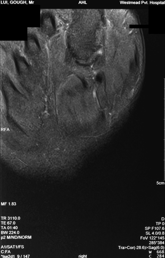
Image Sequence No. 7 – Not too sure – looking at the tissues of the ankle?

Image Sequence No. 8 – Appears to be scan focusing on bones of the ankle joint in detail, from the heel to the front of the foot (maybe).
Conclusion
It was a good day at Advanced Imaging Westmead. Their staff was quick and efficient, and their dedication to operate on Sundays was much appreciated. We await the radiologist’s report, although I suspect the ankle may look just fine. The scans themselves aren’t much fun when they’re “on film” – it takes a bit of imagination and visualization to make it come alive. The animation definitely helps in that regard.
Of course, as an engineer, I’m completely in awe that we (as a species) have achieved so much technologically. The MRI machine is definitely a wonder to behold.
Hi. I like your blog. Quirky, but that’s good. I am an engineer too, interested in almost anything odd.
Apparently the MRIs use big transmitter valves as the voltages etc are beyond semiconductors. They change them routinely and used ones are on-sold to the likes of radio amateurs, as there is still plenty of life left and they are expensive,
Medical imaging has advanced enormously in the last couple of decades, mainly due to computers.
Google my call sign for my blog: VK4ZXI
Regards Drew
Thanks for your comment, and likewise, I just checked out your blog. I’ll be keeping an eye on it in the future. Have fun with the TBS6925 when it gets to you :)!
– Gough
I thought number 6 was a brain scan for a moment! (So much for my hopes of becoming a doctor!)